

Blog
Neurology and Medical Imaging: What You Need to Know
Medical imaging, scanning, and the use of technology are integral parts of how we identify and diagnose neurological conditions today. The proper scan can make all the difference in pinpointing the cause of a patient’s symptoms, paving the way to recovery.
How Medical Imaging Works
With technology, doctors today can gain valuable insights into patients’ bodies to diagnose, monitor, and treat various conditions. The type of scan and the technology that drives it varies similarly to the patient and the area of the body assessed.
MRI Scans
Also known as magnetic resonance imaging, are one of the most common scans used to better look into the brain and soft tissues within the body. This scan is proficient at providing detailed images of the brain, spinal cord, and soft tissues, all while being non-evasive and relatively quick. One of the most notable benefits of the MRI scan is that there is a zero-radiation risk. Instead, MRI scans use a large magnet and computer technology to provide detailed images of the body’s internal system.
This scan also efficiently detects abnormalities such as stroke, tumors, nerve damage, and traumatic brain injury. Occasionally, contrast must be utilized to differentiate between scar tissue and abnormal structures. For example, following spinal surgery, it is often helpful to perform a spinal scan with and without contrast.
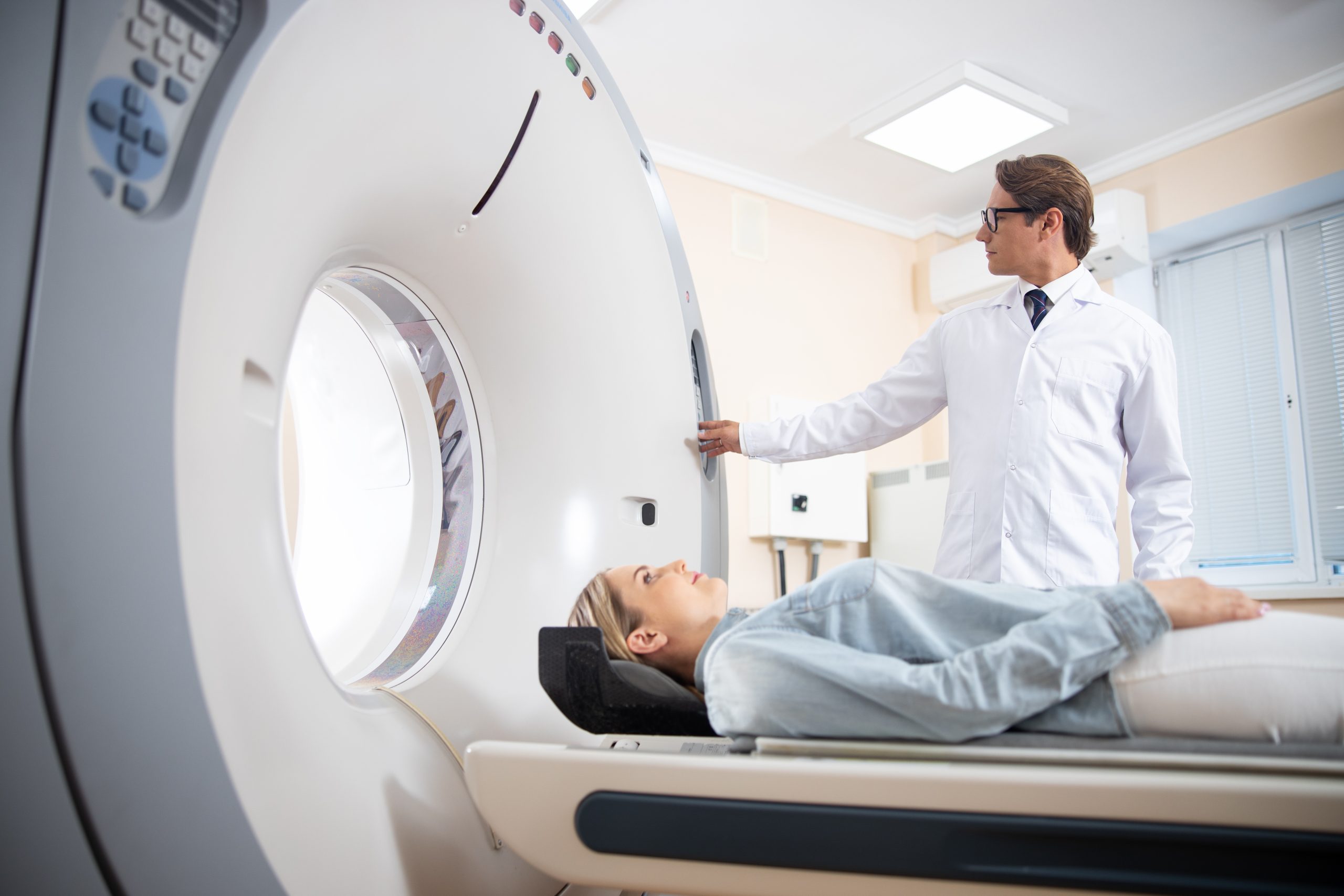
CT Scan
Also known as computerized tomography, is the most powerful X-ray technology available today. A CT Scan identifies and creates images of the body’s soft tissues, bones, and blood vessels. Because of the higher use of radiation, CT scans can produce a far more detailed product than their x-ray counterpart.
The benefit of this scan is that it works efficiently at detecting detailed imaging of internal bleeding and trauma to the bones and soft tissue.
EMG Scan
Also known as electromyography, uses an EMG machine to detect electric activity within the muscles. EMG is beneficial in spotting and diagnosing nerve dysfunction or any disruption in nerve-to-muscle transmission. This type of scan is slightly more invasive, as recording electrodes are inserted into the muscle to translate signals.
The sooner your doctor diagnoses any nerve damage or disruption, the better opportunity to receive proper treatment and reverse or limit the damage.
PET Scan
Also known as positron emission tomography, is one of the most sensitive tests to diagnose brain disorders.
Like the CT scan, a PET scan can provide detailed images of the body. The significant difference between the two is that a PET scan works alongside radioactive chemicals injected into the body to provide precise and detailed imaging.
SPECT Imaging
Also known as single-photon emission computerized tomography, is a test that has become more widely used today. This technology uses nuclear images to provide a detailed image of the brain, heart, and organs.
SPECT tests can detect blood flow and movement within the brain and blood vessels. These images are beneficial in determining clogged vessels and arteries and diagnosing seizure activity within the brain.
FAQs: The Benefits & Risks of Neurological Medical Imaging
Scans make me nervous. Are there any alternatives?
When it comes to medical imaging, many of the scans available today have replaced invasive diagnoses and surgeries. If your doctor or neurologist orders a scan, the goal is to gain insights into the body’s internal condition, which is unavailable to the naked eye. Rest assured, most scans are safe and minimally invasive. However, if your medical condition prevents this, seeking alternative solutions is an option.
Who should avoid medical imaging?
Because every patient and situation is unique, the appropriate use of technology according to the specific condition is required. For example, if you are pregnant or live with a chronic condition, it is best to consult your doctor in detail before proceeding with medical imaging. Patients with implanted medical devices, such as a pacemaker, stimulator, or infusion device, should generally avoid MRI scans. (However, newer devices may be MRI-safe and MRI-compatible) In addition, contrast dyes in scans such as CT and x-ray are usually safe; however, there is a slight risk of allergic reaction and leakage. Consult your doctor if you have any concerns.
I am concerned about radiation exposure. Will this affect my condition?
As scanning technology becomes increasingly advanced, radiation exposure has been a growing topic of concern. For the more basic imaging, such as bone density tests and x-rays, the amount of exposure is relatively minimal and not a cause for concern. For imaging such as CT scans and nuclear imaging (which use a fairly high radiation dose), consulting your doctor will be the best course of action.
One way to navigate radiation risks is to keep track of x-ray history, spanning out scan frequency and asking for a lower dose option. For some, seeking out alternative measures altogether is the best option.
A message from Dr. Kandel
“While imaging has advanced tremendously over the decades, it is still important to remember that it is routinely ordered to confirm a suspected diagnosis or rule out a worrisome or dangerous one. Using today’s modern technology to complement the history and physical examination provides the best possible combined approach to healthcare. Diagnostic testing is just one tool to help you achieve your ultimate goal… improved health!”
Feel free to share this with the people in your life who may benefit from this information! For more insights on neurology, check out our weekly tips on our Neurology Office Facebook page.
“To Cure Sometimes, To Heal Often, To Comfort Always”
Neurology Office, Joseph Kandel M.D. and Associates
“Concierge medicine without the concierge price”